
8 Mitosis and Meiosis.
The processes by which cells divide are complicated and there are a lot of details to remember. Many questions often arise when learning these processes. How many chromosomes are involved in each process? Where did the chromosomes come from; what happens to them; how does it happen; and why is it necessary for all these things to take place? And, perhaps most importantly, how can I remember all this stuff? This lab is designed to make sense out of eukaryotic cell division. If you are having trouble with these processes, this lab should help. In lab, you’ll model these processes with models and practice using the necessary terms to keep track of chromosomes! To prepare, you’ll read the general information on how these processes work so you can come to lab ready to apply your knowledge and see mitosis under the microscope.
The cell cycle
All cells go through the life stages that we call the cell cycle. The first stage is called interphase. During interphase, cells are going through their normal daily cell routines. Liver cells are detoxifying the blood, nerve cells are sending and receiving electrical impulses, skin cells are forming a barrier to the outside world and protecting your inside bits and pieces. Most cells in your body (and those of all other living things) spend most of their lives in interphase. Interphase consists of three phases of its own; the cleverly named G1 (growth 1), S (synthesis), and G2 (growth 2) phases.
During G1, cells have often just come out of cell division and need to replace organelles, structural components, and energy stores used and lost during that process. G1 is the primary growth phase in which these things are replaced. Once the cell has replaced its parts and built up its energy reserves, the DNA is copied. This takes place in the S phase of interphase. Within the nucleus, enzymes unravel the chromosomes and separate the parent DNA strands (existing, complementary DNA molecules) from each other. Other enzymes use the parent DNA strands as templates and build new daughter strands of DNA by attaching nucleotides and reforming the DNA helix (“spiral staircase”).
Once each chromosome is duplicated, cells enter into the G2 phase. During G2, cells get ready to divide. Necessary resources like protein fibers and microtubules are built, and the mitochondria replicate and divide (we’re about to need a whole bunch of energy!). When the cell is ready, it exits Interphase and starts the second stage in the cell cycle.
The second stage involves division of the nucleus and all of the genetic material. This stage takes place in one of two processes, depending on the cell type. Most cells undergo mitosis. Mitosis leads to daughter cells that are genetically identical to the original parent cell and to each other. You might call them clones. Somatic (body) cells in plants, animals, and fungi, and most single-celled protists all go through mitosis. Some cells (those used during sexual reproduction and some really cool, single-celled protist life stages) go through meiosis. Meiosis involves a shuffling of the DNA copies and leads to daughter cells that are genetically unique from each other and the original parent cell. Once the nucleus is divided and all the DNA copies are separated, cells enter the third stage of the cell cycle: cytokinesis.
Cytokinesis is the division of the cytoplasm to create two separate daughter cells. Cytokinesis in animals involves contraction of actin protein filaments causing the plasma membrane to pinch together. This forms a cleavage furrow in the dividing cell. As the filaments continue to contract, the plasma membrane fuses together and forms two new daughter cells out of the original parent cell. Each cell is complete with its own nucleus and set of organelles, and starts the whole cycle over again. In plants, instead of a cleavage furrow, a new cell wall begins to form between the developing daughter cells. This is called a cell plate until it reaches the existing cell walls and completely divides the cells.
So, that’s the basic process that all eukaryotic cells (or most, anyway) go through during their lives. Now let’s look at mitosis and meiosis in more detail.
Mitosis
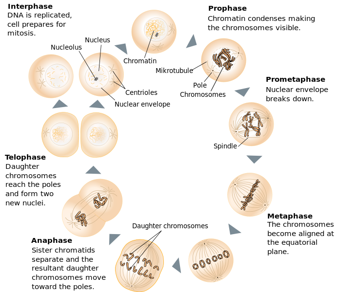
Mitosis comprises four phases: prophase, metaphase, anaphase, and telophase. During prophase, unraveled chromosomes start to condense and coalesce into discrete visible units (what we typically think of as chromosomes). At the same time, the nuclear envelope starts to break down, exposing the chromosomes to the cytoplasm of the cell. The centrosomes are two organelles that are responsible for production and alignment of microtubules (pole-like structures made of tubulin proteins). These two organelles contain the centrioles that aid in this process. While the chromosomes are condensing and the nuclear envelope is dissolving, the centrosomes migrate to opposite poles (ends) of the cell, and start producing a bunch of microtubules. These microtubules radiate out from the centrosomes and form the mitotic spindle. Some of these microtubules (kinetochore microtubules) reach out and bind to each chromosome at protein disks called kinetochores. Kinetochores are located on each side of the centromere of each chromosome. Other microtubules meet each other across the cell (polar microtubules), or radiate out (astral microtubules) to the plasma membrane.
The spindle fibers start to elongate or contract, pushing and pulling the chromosomes toward the middle of the cell. When the chromosomes are all lined up along the middle of the cell, we call this stage metaphase. At this point, all of the centromeres of every chromosome are held on either side by the spindle fibers.
The kinetochore spindle fibers start to contract and tug at the centromere of each chromosome. Enzymes break the protein bonds that hold the centromeres of sister chromatids together, and the sister chromatids separate. As the microtubules retract, the sister chromatids are pulled toward opposite poles of the cell. This stage of nuclear division is called anaphase.
During the last stage of mitosis, telophase, the individual chromatids are all pulled to two centralized locations on either side of the cell. Once there, a new nuclear envelope starts to form around each set of chromatids. As this happens, the chromatids (each is now an individual, separate chromosome) start to unravel within the new nuclei. At the same time, astral and polar microtubules elongate the cell, preparing for division of the cytoplasm during cytokinesis.
While it seems like a lot of information, the phases of mitosis are not hard to remember if you look at the root of each word. “Pro” means first or before, and prophase is the first phase of mitosis. “Meta” means middle, and during metaphase, the chromosomes line up in the middle of the cell. “Ana” means apart or separate, and the sister chromatids are pulled apart during anaphase. “Telo” means end, and telophase is the final phase of mitosis. Also, during telophase, the new nuclei are formed at opposite ends of the cell.
Mitosis modeling
Pair with a teammate and use the chromosome models to work through the stages of mitosis.
- Prepare two pairs of homologous chromosomes: one pair will contain long chromosomes, and the other pair will contain short chromosomes.
- During this exercise, you will model DNA replication, so you will need four sister chromatids representing the long chromosomes, and four sister chromatids representing the short chromosomes.
- Each pair of chromatids should match colors; maternal and paternal copies are different colors.
- In this exercise, the following supplies will be provided to you:
- Magnets = centromeres
- Pipe cleaners = microtubules
- Tape = plasma membranes
- Ribbon/rope =nuclear envelope. Each pair is given two pieces of ribbon so that they can model nuclear envelope disintegration.
- Pipe cleaner wrapped up into a ball= centrosome (microtubules will radiate outward)
- Begin by modeling one cell just after cytokinesis has taken place. Consider how many sister chromatids are present in your simulated cell. Follow your instructor as the class works through the stages of mitosis and complete the questions in your lab workbook to demonstrate your understanding of each phase.
Whitefish microscopy
Obtain a prepared slide of a whitefish blastula and find at least one cell in interphase and in each stage of mitosis: prophase, metaphase, anaphase, and telophase. Draw a cell in each of the stages in Figure 8.2.
Onion microscopy
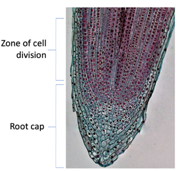
Obtain a prepared slide of a longitudinal section of onion root. Find the zone of cell division and count 100 cells, classifying each into either interphase or one of the phases of mitosis. Record how many cells are in each phase in Table 8.1, and then write your counts on the board. We will use the class data to examine the amount of time individual cells spend in each phase of the cell cycle.
Meiosis
Meiosis is a little more complicated, but it has a lot in common with mitosis. Both processes make use of the terminology: prophase, metaphase, anaphase, and telophase, but meiosis goes through this process twice. This difference leads to one of the most important distinctions between mitosis and meiosis; meiosis is a reduction division process. That means that during cell division, there is a reduction of the total number of chromosomes in the daughter cells. In cells that are going to be used during sexual reproduction (gametes), the number of chromosomes needs to be divided in half. This is important because when two gametes (sperm and ova) join together (fertilization) to form the first cell of a new offspring (called a zygote), half of the offspring’s chromosomes come from each parent. By each parent donating only half of their own genome, the total number of chromosomes in the zygote is the same as the number in each parent. Without this reduction, every generation would double its chromosome count.
Meiosis is divided into two sets of processes, the cleverly named meiosis 1 and meiosis 2. Each of these is further divided into prophase, metaphase, anaphase, and telophase.
During prophase 1, just like in mitosis, the chromosomes condense, the nuclear envelope breaks down, the centrosomes migrate to opposite poles, and the spindle is formed. However, during prophase 1 of meiosis, a very important thing happens. Homologous chromosomes pair together in a process called synapsis and form a protein and chromosome complex called the synaptonemal complex. While in this complex, the chromatids are also called tetrads (tetra means four; there are four chromatids in each complex). While in this state, non-sister chromatids exchange parts. This process is very complex, involves lots of enzymes and proteins, is pretty darn cool, and is called crossing over. Crossing over can happen once or many times between the chromatids of each homologue. At the end of prophase 1, each chromatid is a unique combination of the original homologous chromosome materials. While all of this is going on, spindle fibers connect to the paired chromosomes and begin to move them toward the middle of the cell.
In metaphase 1, the synaptonemal complex of each homologous pair is lined up along the central plane of the cell. The alignment of these homologues is completely random, such that the original source (paternal, maternal) homologue can line up on one side or the other of the central plane, without regard to the alignment of the other homologous pairs. It is this random alignment of parental homologues that provides the basis for the genetic concept of independent assortment. Independent assortment basically states that offspring inherit physical traits from their parents without regard to the forms of other traits that they might inherit. In other words, inheritance of each trait happens independently from inheritance of any other traits.
During anaphase 1, the synaptonemal complexes break down, and homologues are pulled to opposite poles of the cell. It’s important to note that sister chromatids remain together in this process, and that one of each homologue (now consisting of a recombination of the parental chromosomes) is going to end up in each daughter cell.
During telophase 1, a new nuclear envelope forms around each set of chromosomes, the spindle elongates the cell, and the cell prepares to divide.
Cytokinesis takes place between meiosis 1 and meiosis 2 resulting in daughter cells that have half the number of chromosomes as the parent cells. This is the reduction part of meiosis. In diploid organisms (those with pairs of homologues; humans, for example) the daughter cells after meiosis 1 are haploid, they have a single representative chromosome from each homologous pair. It is important to note that although the cells have half the total number of chromosomes as the parent cell, each chromosome still consists of two sister chromatids. Because of crossing over during prophase 1, these sister chromatids are recombinant and are no longer genetically identical.
Depending on the particular organism, the time between meiosis 1 and 2 can be brief or very long. There may also be a brief interphase-like stage during which the cell replaces organelles and energy stores before continuing on to meiosis 2. The combined cytokinesis and pre-meiosis 2 prep phase is sometimes called interkinesis. Interkinesis differs from interphase in the fact that there is no DNA replication between meiosis 1 and 2.
Meiosis produces genetically unique daughter cells. Homologous chromosomes separate in meiosis 1 and sister chromatids separate during meiosis 2. Meiosis image from OpenStax, CC-BY.
Meiosis 2 is a division of the haploid daughter cells from meiosis 1. In prophase 2, the nuclear envelope breaks down, the spindle forms, and chromosomes are moved toward the middle of the cell. In metaphase 2, the chromosomes line up along the central plane of the cell. In anaphase 2, sister chromatids are separated and pulled to opposite poles of the cell. In telophase 2, new nuclear envelopes form around each chromosome group, and during cytokinesis the cell divides. The combined result of meiosis 1 and 2 is four daughter cells from one parent cell. Each daughter cell contains one homologue (composed of a single chromatid) from each chromosome pair that consists of a unique combination of DNA from the original parent cell.
Depending on the organism, the daughter cells may be further modified to produce gametes in the process of gametogenesis. In mammals, for example, the production of sperm cells in males is called spermatogenesis, and the production of ova cells in females is called oogenesis. By contrast, in some protists the outcome of meiosis is the active haploid stage in which the organisms spend most of their lives. Isn’t biology cool!?
Meiosis modeling
Following your instructor, work through the stages of meiosis using your cell model and answer the questions below.
At the end of the lab, you’ll complete more review questions at the end of the workbook to make sure you can use the terms of cell division accurately. These are complex processes so taking the time to work through each mechanism step by step during lab makes a big difference in your understanding.
